The Cupid Peptide Company
EXPERTS IN CELL PENETRATING PEPTIDE
CUSTOM DESIGN AND MANUFACTURE
Cupid-MDM2-A
Product Description:
Cupid-MDM2-A peptide:
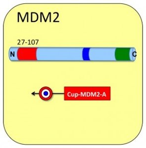
• Cargo: Residues 27 to 107 of human MDM2
• Domain type: SWIB Motif, SWIB / MDM2 domain (P53 binding)
• 81 Amino acids
LVRPKPLLLKLLKSVGAQKDTYTMKEVLFYLGQYIMTK
RLYDEKQQHIVYCSNDLLGDLFGVPSFSVKEHRKIYTM
IYRNL
Product Characteristics:
• Molecular weight (daltons): 17212
• Isoelectric point (pI): 9.6
• Extinction Coefficient (A280 reduced) : 17420
• Solubility : >200 micromolar
• Purity: 83%
• Cell-Permeation : Passes
• 1 Unit = 10 nanomoles = 0.172 mg
Cupid-MDM2-A Peptide Data:
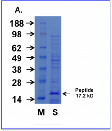
Cell permeating Cupid MDM2 peptide AA. Purified Cupid-MDM2-A peptide was subjected to SDS-PAGE alongside a prestained molecular marker ladder. The gel was then stained for protein with a commercial coomassie-based stain.
M = Weight markers shown in kD
S = Cupid-MDM2-A Peptide sample.
Cell permeating Cupid MDM2 peptide A
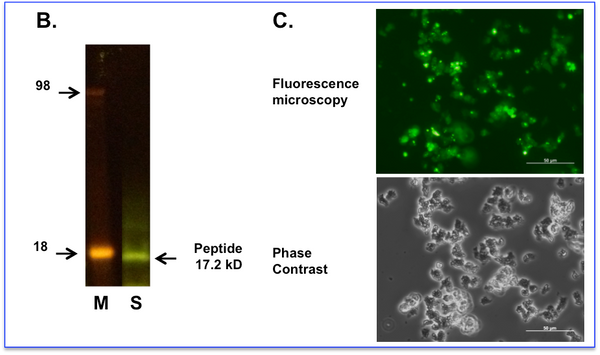
B. Cupid-MDM2-A peptide labelled with fluorescein was subjected to SDS-PAGE and observed with a blue light transilluminator.
M = Weight markers shown in Kd
S = Labelled Cupid-MDM2-A Peptide sample.
C. Labelled peptide was incubated with living cells at 10 micromolar for 60 minutes before exchanging the media and washing the cells. Cells were imaged using a fluorescence microscope with filter sets for Fluorescein (Upper Panel) or phase contrast (Lower Panel). Fluorescent images of treated cells are taken at a setting where the background (autofluorescence) of the untreated cells is at the threshold of detection. We observe the peptide fluorescence distributed diffusely throughout all cells.


